- Blog
Knee Replacement Alternatives
Posted on 07-22-2026 in Knee by Dr. Erik Nilssen
Posted on 07-22-2026 in Knee by Dr. Erik Nilssen
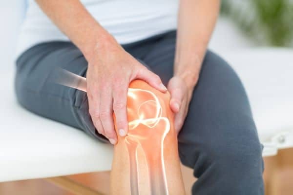
According to the Agency for Healthcare Research and Quality, more than 600,000 knee replacements are performed each year in the United States. As people are developing the loss of cartilage in their knees, the joints start seeing a lot more stress. This comes from the “bone on bone” changes that are occurring. As the bones see more stress, we often start seeing what are called “insufficiency fractures” or bone bruises on MRI scans.
The Orthopedic literature seems to indicate that a lot of the pain from arthritis comes, not from the bone-on-bone changes, but from the developing insufficiency fracture and bone bruising. As alternatives to knee replacement surgery, the following procedures may be suitable pending a variety of factors:
SCP – An Alternative Procedure
Over the last couple of years when we see these bony changes on MRI, we inject a bone graft substitute into the bone to seal up the insufficiency problem. This is called a subchondroplasty.
Think of it as injecting a type of wall grout inside the bone to help support it while it attempts to heal.
For those patients who are too young, too obese, or not interested in a total knee replacement, this minimally invasive procedure could be a pain-reducing procedure.
SCP – Early Results
The results are early but are quite promising. You may be the right patient for this procedure if your pain seems greater than you would think from the amount of arthritis present, or the tenderness is actually on the bone rather than in the joint.
Please see below additional details about the SCP Procedure.
KNEE SUBCHONDROPLASTY
Bone Marrow Lesions (BML) are subchondral bone defects visible on an MRI. You can only see them on fat-suppressed MRI sequences (PDFS, T2FS, etc.) where they look like a hazy white spot against the darker bone background. According to pathologists, BML represents a trauma-related healing response like subchondral bone micro-trabecular fractures. They can be found in any bone that sees weight-bearing or repetitive motion stress.
In these patients, present guidelines for treatment suggest an initial course of conservative care to help heal the defect. Then, the Subchondroplasty® (SCP®) Procedure might be an option if the defect remains.
WHAT IS THE SUBCHONDROPLASTY PROCEDURE?
This procedure is a fluoroscopically-assisted, minimally-invasive procedure surgeons use for targeting and filling subchondral bone defects, such as BML, with AccuFill® PF Bone Substitute Material (BSM). This is a biomimetic, hard-setting bone substitute.
Unlike standard “knee replacement surgery” where the surgeon removes the damaged or diseased knee joint and replaces it with an artificial one, the Subchondroplasty Procedure is a minimally-invasive procedure surgeons use to delay or prevent knee osteoarthritis progression by treating the underlying causes of osteoarthritis knee mobility limitations and pain. The term “subchondroplasty” literally refers to “surgical repair below the cartilage.”
The Subchondroplasty Procedure:
Despite physical examination and negative X-rays when BMLs are causing the pain, the Subchondroplasty Procedure could ease the pain.
The surgeon performs Subchondroplasty along with arthroscopy of the impacted knee for:
WHO IS AN IDEAL CANDIDATE FOR THIS PROCEDURE?
You might be a candidate for Subchondroplasty if you have:
Frequently, osteoarthritis is the cause of knee pain and is the most common type of arthritis. Because it’s a “wear-and-tear condition” occurring as your joints degenerate with age and use, it’s aptly named degenerative arthritis. Traditionally, doctors diagnose knee osteoarthritis by evaluating:
The affected joints’ X-rays will have a characteristic appearance where the ends of the bones look closer to one another because of joint space narrowing with the wearing away of cartilage.
Additionally, fluid-filled cavities or cysts might be noticeable in the bone as your body reacts to cartilage destruction. The doctor might observe uneven joints or increased bone density since when cartilage doesn’t cushion bones any longer, the bones rub against each other with more pressure and create pain and friction. Your body reacts to this increased pressured force by increasing bone density and laying down more bone which creates bone spurs (osteophytes) and uneven joint surfaces around the joint margins.
According to a study published by the National Institutes of Health, there’s a 45% lifetime risk of developing knee osteoarthritis. Many individuals expecting more active lifestyle years are searching for alternatives to the full knee replacement procedure which is invasive and comes with a lengthy recovery period. It also comes with certain lifetime activity limitations (no jumping/running for instance). Subchondroplasty provides them with such an option.
THE SURGICAL PROCESS
1. Preoperative Plan: Find the BML bone defect through a fat-suppressed MRI scan; plan trajectory and approach based on the location of the defect.
2. Target the Bone Defect: Using intraoperative fluoroscopy and arthroscopy, localize the bone defect in relation to the findings on the MRI.
3. Access the Defect: Drill the proper AccuPort® Delivery Cannula to reach the bone defect.
4. Fill the Bone Defect: Inject the subchondral bone defect with AccuFill BSM.
The Subchondroplasty Procedure will target and fill the BMLs in those not responding to conservative treatments.
Your surgeon will use AccuFill® Injectable Bone Substitute Material. This is a self-setting, osteoconductive, macroporous, calcium phosphate, injectable bone graft substitute material made for filling bony gaps or voids of the skeletal system of the spine, pelvis, and extremities that aren’t intrinsic to the bony structure’s stability.
The defects might be traumatic injury-related osseous defects or surgically created osseous defects to the bone. The AccuFill injectable resorbs and is replaced by new bone during the process of healing.
You will likely experience some discomfort and pain in the surgery area for a day or two after your procedure. The doctor may prescribe you pain medicine to manage it. They will recommend crutches for one to two weeks after your procedure to reduce weight-bearing on your operated leg. They may also recommend physical therapy to regain mobility and strength in your knee.

Have you ever taken a step and felt like there was a pebble stuck in your shoe, only to stop, check, and find nothing there? It's a surprisingly common complaint. Many patients describe the sensation as though they're walking on a small rock, a wrinkle in their sock, or something that simply won't go away. While it may seem like a minor annoyance at first, persistent pain in the ball of the foot is often your body's way of signaling that something isn't functioning as it should. Because several conditions can cause similar symptoms, understanding what may be behind the discomfort is the first step toward finding lasting relief.
For many patients living with chronic low back pain (LBP) and advanced knee arthritis, there comes a point when the treatments that once provided relief no longer seem to work as well. Medications may offer only temporary comfort, injections may become less effective over time, and everyday activities like walking, climbing stairs, or even getting out of a chair can become increasingly difficult. At that point, many patients ask the same question: "Am I ready for surgery, or are there other options I should consider first?" The answer is rarely as simple as "yes" or "no."

May is Arthritis Awareness Month, an opportunity to increase public understanding of arthritis and its impact on millions of lives. Established by the Arthritis Foundation, this national observance highlights the importance of early diagnosis, effective treatment, and ongoing research to improve the quality of life for those with arthritis.